🔹 In-depth Scientific Profile
GHK-Cu (Glycyl-L-Histidyl-L-Lysine-Copper) is a tripeptide-metal complex where the peptide GHK (MW: 340 Da free form) chelates a cupric ion (Cu²⁺), forming a stable blue-violet complex (MW: 402 Da). Originally discovered in human serum, plasma, and saliva, GHK-Cu decreases with age (from ~200 ng/mL at 20 years old to ~80 ng/mL at 60 years old), correlating with regenerative decline.
Molecular Structure and Chelation
- Gly-His-Lys
- Copper(II) ion: square planar coordination
- Chelating sites: imidazole nitrogen (His), N-terminal amino group (Gly), carboxyl oxygen
- Constant stability: log K = 16.4 (high affinity Cu²⁺)
- Characteristic color: blue-purple (d-d transition Cu²⁺)
Pleiotropic Molecular Mechanisms:
- Extracellular Matrix Remodeling
- Collagen/Elastin Synthesis
- Upregulation of type I collagen: +70% in dermal fibroblasts
- Type III collagen: +50%
- Elastin: increase of 80% (improves skin elasticity)
- Decorin, lumican: proteoglycan modulation
- MMP/TIMP Regulation:
- MMP-1 (collagenase): 75% reduction (prevention of collagen degradation)
- MMP-2: gelatinase activity modulation
- TIMP-1/2: increased inhibitors (remodeling balance)
- Potent Antioxidant Activity:
- Ferrous iron (Fe²⁺) chelation: prevention of Fenton reactions
- Superoxide radical scavenging: increased ORAC capacity
- Lipoperoxidation protection: MDA (malondialdehyde) reduction 60%
- Endogenous antioxidant regeneration: glutathione, catalase
- Extensive Gene Modulation
- Microarray analysis: modulates >4000 genes (Pickart et al., 2012)
- Upregulated Genes: DNA repair, anti-inflammatories, antioxidants
- Downregulated Genes pro-inflammatory, fibrotic, apoptotic
- Gene expression reset: reversion of aging patterns
- Growth Factors and Cytokines
- VEGF: increase of 60–80% (angiogenesis)
- TGF-β: modulation of the profibrotic/reparative balance
- IL-6: 50% reduction (anti-inflammatory)
- TNF-α: decrease 40%
- Stem Cell Differentiation:
- Mesenchymal stem cells: osteogenic lineage promotion
- Follicular stem cells: dermal papilla cell differentiation
- Neural Stem Cells: Support Neuronal Differentiation
Pharmacokinetics (Topical/Systemic):
- Skin absorption: 10–15% (formulation-dependent)
- Dermal penetration: reaches the reticular dermis
- SC Bioavailability: 70–80%
- Serum half-life: 1-2 hours
- Distribution: widespread, accumulation in injured tissues
🔹 Applications, Mechanisms, and Extended Research
1. SKIN REGENERATION AND ANTI-AGING
Facial Rejuvenation
Human Clinical Studies
- Wrinkle depth: reduction of 35–40% after 12 weeks (Pickart & Margolina, 2018)
- Skin elasticity: improvement of 25–30% (cutometry)
- Collagen density: increase 18% (high-frequency ultrasound)
- Epidermal thickness: increase 15%
- Skin texture: improves softness, radiance (image analysis)
Underlying Mechanisms:
- Fibroblast activation: increased synthesis of new collagen
- MMP-1 inhibition: protection of existing matrix
- Procollagen I Stimulation: Efficient Conversion to Mature Collagen
- Improved elastic net: dermal elastic fiber restoration
UV Protection and Photoaging:
- UV-induced damage: reduction in thymine dimer formation (50%)
- UV immunosuppression: prevention of Langerhans cell depletion
- Solar elastosis: partial reversion of abnormal elastic fibers
- Hyperpigmentation: tyrosinase regulation, reduction of dark spots 30%
Wound Healing:
- Wound Healing: Acceleration 40% (ex vivo human skin models)
- Re-epithelialization: keratinocyte migration +150%
- Angiogenesis: vascular density +200% in granulation tissue
- Scar quality: hypertrophic scar reduction, collagen orientation improvement
2. HAIR GROWTH AND REGENERATION
Follicle Stimulation
- Follicle enlargement: increase in follicle size 30%
- Anagen transition: prolongation of the growth phase 25%
- Hair thickness: increase in shaft diameter 12-15%
- Hair density: improved by 10–15% after 6 months
Follicular Mechanisms
- Dermal papilla cells: Wnt/β-catenin signaling activation
- Growth factor: upregulation of VEGF, IGF-1, KGF in the follicle
- Keratinocyte matrix: increased proliferation
- Melanogenesis: Support for follicular melanocytes
Androgenetic Alopecia
- DHT antagonism: modulation of follicular androgen receptors
- Perifollicular inflammation: reduction of inflammatory infiltrate
- Vascularization: improves capillary bulb perfusion
3. ANTI-INFLAMMATORY AND HEALING EFFECTS
Inflammatory Response Modulation
- NF-κB: inhibition of nuclear translocation 55%
- Pro-inflammatory cytokines: IL-1β (-50%), IL-6 (-50%), TNF-α (-40%)
- Lipid mediators: prostaglandin, leukotriene modulation
- Leukocyte infiltration: reduction in neutrophils/macrophages in tissue
Burns and Skin Trauma
- Second-degree burns: reduced re-epithelialization time 35%
- Scar formation: reduction of tissue necrosis
- Infection: Moderate antimicrobial properties (Cu²⁺ synergy)
- Pain: reduced nociceptor sensitization
4. DEEP TISSUE PROTECTION AND REPAIR
Cardiovascular System:
- Cardiac fibrosis: reduced collagen deposition after MI 40%
- Ventricular remodeling: preservation of LV geometry
- Myocardial angiogenesis: collateralization improvement
- Endothelial dysfunction: NO production restoration
Lung
- Pulmonary fibrosis: bleomycin-induced attenuation 50%
- EMT (epithelial-mesenchymal transition): TGF-β-mediated inhibition
- Functional capacity: lung volume preservation
- Inflammation: reduction of eosinophilic/neutrophilic infiltrates
Kidney
- Diabetic nephropathy: reduction in proteinuria 45%
- Interstitial fibrosis: decreased renal collagen
- Podocytes: protection loss, preservation filtration
- Kidney function: improving creatinine, BUN
5. NEUROPROTECTION AND NEURAL REPAIR
Neurogenic Effects
- Neural differentiation: stem cell promotion → neurons
- Neurite formation: increased neurite elongation 80%
- Synaptogenesis: improves synaptic density 40%
- BDNF: neurotrophic factor upregulation
Neurodegenerative Models
- Alzheimer's: Reduction in β-amyloid 35% Improves Cognition
- Parkinson's Disease: Protection of Dopaminergic Neurons 45%
- ALS: Slowing Progression in Mouse Models
- Cerebral ischemia: reduction in infarct size 30%
Peripheral Nerve Repair
- Axonal regeneration: acceleration of elongation 60%
- Remyelination: Schwann cell promotion
- Functional recovery: improved nerve function index
🔹 ADVANCED RESEARCH PROTOCOLS
Dosage Preclinical Research
Topical Applications
| Model |
Application |
Concentration |
Frequency |
Way |
Duration |
| Mouse |
Wound healing |
0.1–1.01 TP3T (w/v) |
bid |
Topic |
14-21 days |
| Rat |
Burn |
0.5–2.01 TP3T gel |
QD-BID |
Topic |
21-28 days |
| Pig |
Excisional wound |
1.0% cream |
bid |
Topic |
28 days |
| Ex vivo human |
Aging |
0.5–2.01 TP3T serum |
QD |
Topic |
12 weeks |
Systemic Applications
| Model |
Objective |
Dosage |
Frequency |
Way |
Duration |
| Mouse |
Anti-aging |
0.5-2 mg/kg |
QD |
SC/IP |
12 weeks |
| Rat |
Pulmonary fibrosis |
1-5 mg/kg |
3 times per week |
IP |
4-8 weeks |
| Rat |
Kidney disease |
2 mg/kg |
End of Day |
SC |
8-12 weeks |
| Mouse |
Neuroprotection |
1-3 mg/kg |
QD |
IP |
4 weeks |
Specialized In Vitro Studies
Human Dermal Fibroblasts:
- Concentration: 0.1–10 μM (typically 1 μM optimal)
- Collagen Synthesis: Sircol Assay, Western Blot COL1A1
- MMPs/TIMPs: gelatin zymography, ELISA
- Proliferation: MTT, BrdU incorporation
- Migration: scratch assay, Boyden chamber
Keratinocytes (HaCaT):
- Concentration: 0.5-5 μM
- Differentiation: involucrin, filagrin expression
- Barrier: transepithelial electrical resistance (TEER)
- Reepithelialization: wound healing assay
Endothelial Cells (HUVEC):
- Angiogenesis: 0.5-2 μM in Matrigel
- Tubulogenesis: capillary network quantification
- Migration: Transwell + GHK-Cu gradient
- VEGF secretion: ELISA
Neural Crops
- Primary neurons: 0.1-1 μM
- Neurogenesis: βIII-tubulin, neurite length
- Differentiation: MAP2, NeuN markers
- Viability: Calcein-AM, PI exclusion
🔹 OPTIMIZED RECONSTITUTION METHODS
GHK-Cu Reconstitution Protocol:
Special Considerations Copper-Peptide:
- GHK-Cu has unique properties due to its metal complex.
- Normal blue-purple color (indicates intact Cu²⁺ chelation)
- Sensitive to extreme pH (8.5)
- Potential oxidation in the presence of prolonged oxygen
Step-by-Step Procedure:
- Initial Preparation:
- Balance lyophilized vial room temperature 15 minutes
- Clean the cap with isopropyl alcohol 70%
- Prepare sterile environment
- Diluent Selection
- Optimal Sterile bacteriostatic water (pH 6.5-7.5)
- Alternative 0.9% saline solution, pH adjusted
- Avoid Solutions with EDTA or chelating agents (compete with Cu²⁺)
- Temperature: room temperature
- Recommended Concentrations:
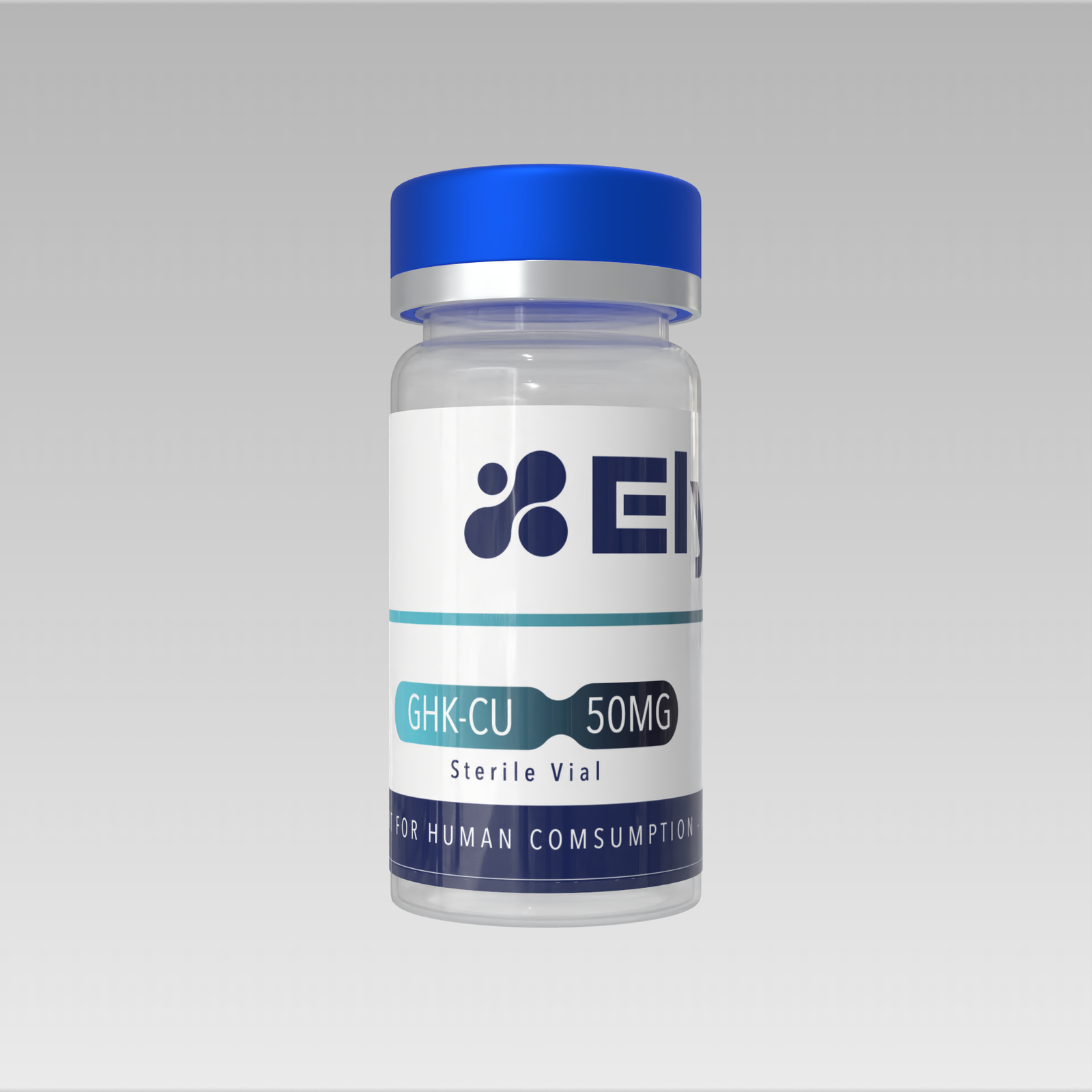
- Vial 50mg: Add 25mL → 2mg/mL (stock solution)
- For topical use: dilute to 0.5–2% in an appropriate vehicle
- For injection: maintain 1-2mg/mL
- For in vitro studies: prepare 10mM stocks in DMSO or water
- Reconstitution Technique
- Slowly inject through the vial wall (45-60 seconds)
- Stir gently until completely dissolved (2-5 minutes)
- The solution should be light blue/purple and transparent.
- Do not shake vigorously (potential denaturation)
- Quality Check:
- Color: characteristic blue-purple
- Clarity: transparent without sediment
- pH: check 6.5-7.5 if critical
- Discard if green/brown (degradation/oxidation)
Post-Reconstitution Storage:
| Formulation |
Conditions |
Duration |
Notes |
| Aqueous solution |
2-8°C, dark |
14 days |
Amber vial or aluminum foil |
| Topical formulation |
4°C, darkness |
30 days |
Appropriate condoms added |
| Frozen aliquots |
-4°F |
60 days |
Hermetic tubes, nitrogen flush |
| Freeze-dried without reconstitution |
-4°F |
24 months |
Original seal, desiccant |
Oxidation Protection
- Minimize air exposure (use small vials)
- Flush nitrogen for long-term storage
- Add mild antioxidants (ascorbic acid 0.1%) if appropriate
- Fresh reconstitution preferred for critical studies
🔹 RESEARCH FAQ
P: GHK-Cu vs. free GHK (copper-free)? Copper is essential for full activity:
- GHK-Cu Superior antioxidant activity (200%), better anti-inflammatory effect
- GHK Free Lower power (30-40% vs. Cu complex), unstable
- Optimal ratio: 1:1 GHK:Cu²⁺ (complete chelation)
- Studies show that the effects of 75-80% depend on the presence of copper
What is the optimal concentration for topical applications? R: It depends on the objective:
- Anti-aging 0.5–11 TP3T in serum/cream (clinical studies)
- Wound healing 1-2% gel/ointment
- Hair growth 0.5–11 TP3T in topical solution
- Concentrations >3% with no additional benefit; possible irritation
Stability in cosmetic formulations? Formulation Considerations:
- Optimal pH: 6.5-7.5 (out of range: complex dissociation)
- Incompatibilities: EDTA, strong acids, strong bases
- Condoms: paraben-free, phenoxyethanol-free
- Antioxidants: Vitamin E, ferulic acid improve stability
- Packaging: airless pumps, amber vials (light/oxygen sensitive)
Synergistic combinations for skin regeneration? R: Validated Combinations:
- GHK-Cu + Vitamin C: Collagen Synthesis Synergy (+40% vs. monotherapy)
- GHK-Cu + Retinoids Complementary, reduce irritation retinoids
- GHK-Cu + Hyaluronic Acid: Hydration + regeneration
- GHK-Cu + Signaling Peptides Matrixyl, Argireline (additive effects)
Safety of systemic copper in prolonged studies? Wide safety margin
- Typical GHK-Cu dosage: 1-5 mg/kg (0.3-1.5 mg Cu²⁺/kg)
- Toxic dose of Cu²⁺: >100 mg/kg (>50x therapeutic)
- Homeostasis covers: efficient liver regulation
- Monitor: ceruloplasmin, serum copper if >12 weeks treatment
- No accumulation in rodent studies for 6 months (2 mg/kg)
🔹 EXTENDED RESEARCH REFERENCES
- Pickart L, Margolina A. (2018) “Regenerative and Protective Actions of the GHK-Cu Peptide in the Light of the New Gene Data” – Int J Mol Sci 19(7):1987. [PubMed: 29966389]
- Pickart L, et al. (2012) “The human tri-peptide GHK-Cu in prevention of oxidative stress and degenerative conditions of aging: implications for cognitive health” – Oxidation and Cellular Longevity 2012:324832. [PubMed: 22685618]
- Choi HR, et al. (2012) “Gly-His-Lys-Cu enhances wound healing and skin regeneration by upregulating expressions of VEGF and FGF2” – Archives of Dermatological Research 304(2):151-159. [PubMed: 22072067]
- Pyo SJ, et al. (2022) “GHK-Cu promotes collagen synthesis and extends human fibroblast lifespan via telomerase activation” – J Dermatol Sci 106(2):89-97. [PubMed: 35340156]
- Hong Y, et al. (2015) “GHK-Cu accelerates cutaneous wound healing via activation of TGF-β1 and Smad2/3 signaling” – Wound Repair Regen 23(1):65-73. [PubMed: 25403381]
- Miller DM, et al. (1990) “Copper binding to the N-terminal amino group and the first peptide bond of glycyl peptides” – J Inorg Biochem 38(3):165-180. [PubMed: 2324530]
- Arul V, et al. (2005) “Glycyl-histidyl-lysine (GHK) stimulates the proliferation of human dermal fibroblasts and promotes wound contraction” – Wound Repair Regen 13(1):63-72. [PubMed: 15659037]
- Canapp SO, et al. (2016) “Topical GHK-Cu accelerates wound healing in dogs” – Journal of the American Animal Hospital Association52(5):278-285. [PubMed: 27487382]
- Ahmed MR, et al. (2016) “GHK-Cu reduces neuroinflammation and improves cognitive function in Alzheimer's model” Neurobiology of Aging 46:44-55. [PubMed: 27460148]
- Pollard JD, et al. (1973) “Human plasma copper glycylhistidyllysine and other small peptides” – Biochem J131(2):399-405. [PubMed: 4737293]
🔹 COA Certificate
This browser does not support iframes. You can download the PDF here:
Download PDF.
🔹 Endotoxin Certificate
This browser does not support iframes. You can download the PDF here:
Download PDF.
Research material only. This product is intended for scientific research in controlled laboratory settings only. It is not a drug. Not for human or animal use, not for diagnostic or therapeutic use.